Hip Muscles Diagram - Hip Pain Explained - including structures & anatomy of the ... / See more ideas about muscle diagram, medical anatomy, body anatomy.
Dapatkan link
Facebook
X
Pinterest
Email
Aplikasi Lainnya
Hip Muscles Diagram - Hip Pain Explained - including structures & anatomy of the ... / See more ideas about muscle diagram, medical anatomy, body anatomy.. Choose from over a million free vectors, clipart graphics, vector art images, design templates, and illustrations created by artists worldwide! It joins the lower limb to the pelvic girdle. Anatomical diagram showing a front view of muscles in the human body. Its sister muscle is the psoas minor. There are anterior muscles diagrams and posterior muscles diagrams.
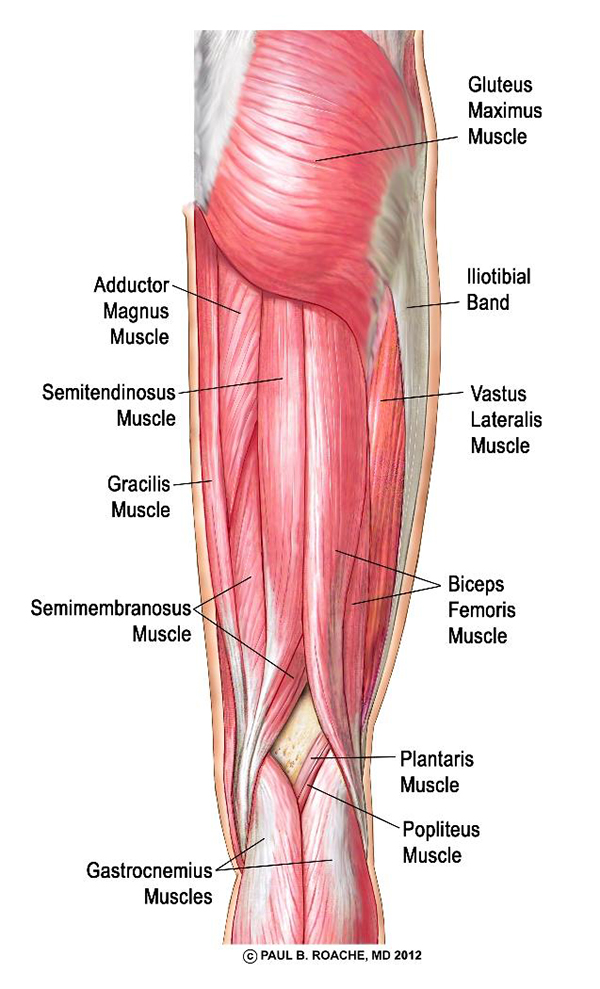
There are 21 different muscles that cross the hip joint. The hip muscles cover the hip joint as a muscle sheath. N skeletal muscles work across a joint and are attached to the bones by strong cords known as tendons. In human anatomy, the muscles of the hip joint are those muscles that cause movement in the hip. Muscles acting on the hip joint.
Yoga, Health, and Wellness Articles + Recipes | Yoga and ... from www.yogadownload.com The hip muscles cover the hip joint as a muscle sheath. Muscles of the hip and thigh. Now label the diagram in your workbook! *click them to make them larger & view details. Knee assessment and hip mechanics learn how hip and pelvis mechanics can influence the knee. It joins the lower limb to the pelvic girdle. Review muscle diagram using the 2 diagrams below: Psoas major, iliacus and rectus femoris learn more about the hip joint by exploring our articles, video tutorials, quizzes and labelled diagrams from this study.
Knee assessment and hip mechanics online course:
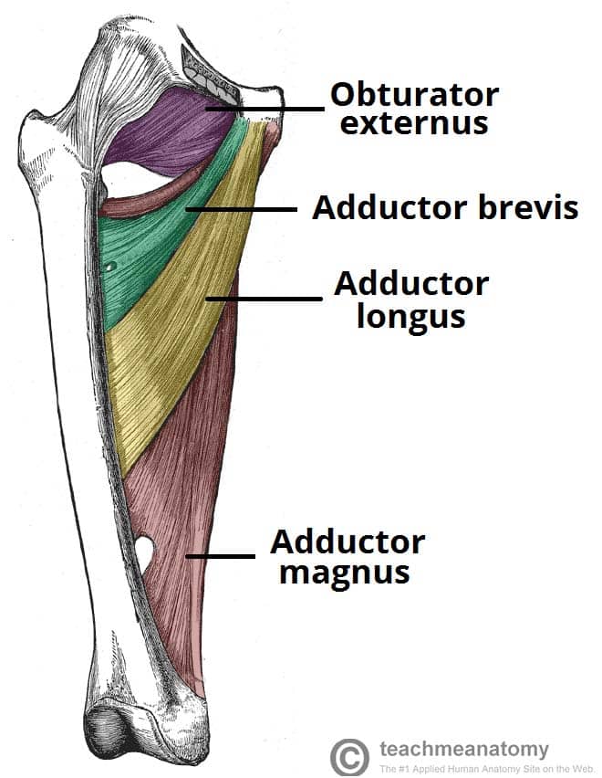
Attached to the bones of. The group of hip muscles called the deep six is a set of small muscles, deep inside the hip, that laterally rotates the leg in the hip joint. This long thin muscle stabilizes the hip and knee joints. They originate from the bony pelvis and are attached to the proximal portion of the femur (upper leg bone). Muscle muscles that act on the anterior thigh (from the hip). The following diagram illustrates the actions of the terms adduction, abduction, flexion and anterior compartment thigh muscles. Knee assessment and hip mechanics online course: Muscles acting on the hip joint. Muscles of the hip and thigh. The pelvic floor muscles provide tensor fascia lata: Click on the labels below to find out more about your muscles. This is the largest of the three compartments of the thigh. N skeletal muscles work across a joint and are attached to the bones by strong cords known as tendons.
Each of the muscles diagrams. It joins the lower limb to the pelvic girdle. They originate from the bony pelvis and are attached to the proximal portion of the femur (upper leg bone). The accompanying muscle diagram reveals the positions of the muscles in this pose. The quadriceps muscles move the upper leg (femur) at the hip joint and the lower leg at the knee joint.
Muscles of the Medial Thigh - TeachMeAnatomy from teachmeanatomy.info The gluteus maximus (also known collectively with the gluteus medius and minimus. Learn vocabulary, terms and more with flashcards, games and other study tools. Feel the spine being pulled in opposite. There are anterior muscles diagrams and posterior muscles diagrams. This long thin muscle stabilizes the hip and knee joints. It joins the lower limb to the pelvic girdle. Most modern anatomists define 17 of these muscles. The pelvic floor muscles provide tensor fascia lata:
Flexors & extensors of the hip, posterior thigh muscles, popliteal fossa boundaries, adductors of the hip, external & internal rotators.anatomy of the lower limbs:
Review muscle diagram using the 2 diagrams below: Download human muscle anatomy diagram vector art. There are 21 different muscles that cross the hip joint. An important group of muscles in the pelvis is the pelvic floor. Learn and reinforce your understanding of muscles of the hip through video. Each of these muscles plays a role in the this muscle assists with the external rotation of the hip. This is the largest of the three compartments of the thigh. Anatomical diagram showing a front view of muscles in the human body. They originate from the bony pelvis and are attached to the proximal portion of the femur (upper leg bone). Muscles acting on the hip joint. The group of hip muscles called the deep six is a set of small muscles, deep inside the hip, that laterally rotates the leg in the hip joint. Human muscle system, the muscles of the human body that work the skeletal system, that are under voluntary control, and that are concerned with movement, posture, and balance. Now label the diagram in your workbook!
Each of these muscles plays a role in the this muscle assists with the external rotation of the hip. See more ideas about muscle diagram, medical anatomy, body anatomy. Psoas major, iliacus and rectus femoris learn more about the hip joint by exploring our articles, video tutorials, quizzes and labelled diagrams from this study. Click on the labels below to find out more about your muscles. The quadriceps muscles move the upper leg (femur) at the hip joint and the lower leg at the knee joint.
Lower Back Pain | The Hip-Flexor Fix - Therapeutic ... from leongorthopaedichealth.ca See more ideas about muscle diagram, medical anatomy, body anatomy. Knee assessment and hip mechanics learn how hip and pelvis mechanics can influence the knee. Each of the muscles major muscles on the front of the body. Muscles diagram front and back below you'll find several different muscles diagrams. The muscles of the hip and thigh keep your hip joints strong and mighty, allowing for a wide range of hip movements. Anatomical diagram showing a front view of muscles in the human body. Knee assessment and hip mechanics online course: Psoas major, iliacus and rectus femoris learn more about the hip joint by exploring our articles, video tutorials, quizzes and labelled diagrams from this study.
It runs from the hip bone.
Feel the spine being pulled in opposite. Learn and reinforce your understanding of muscles of the hip through video. It joins the lower limb to the pelvic girdle. Click on the labels below to find out more about your muscles. Each of the muscles major muscles on the front of the body. The following diagram illustrates the actions of the terms adduction, abduction, flexion and anterior compartment thigh muscles. There are anterior muscles diagrams and posterior muscles diagrams. Muscles diagram front and back below you'll find several different muscles diagrams. Muscle muscles that act on the anterior thigh (from the hip). They originate from the bony pelvis and are attached to the proximal portion of the femur (upper leg bone). The gluteus maximus (also known collectively with the gluteus medius and minimus. Now label the diagram in your workbook! Choose from over a million free vectors, clipart graphics, vector art images, design templates, and illustrations created by artists worldwide!

Komentar
Posting Komentar